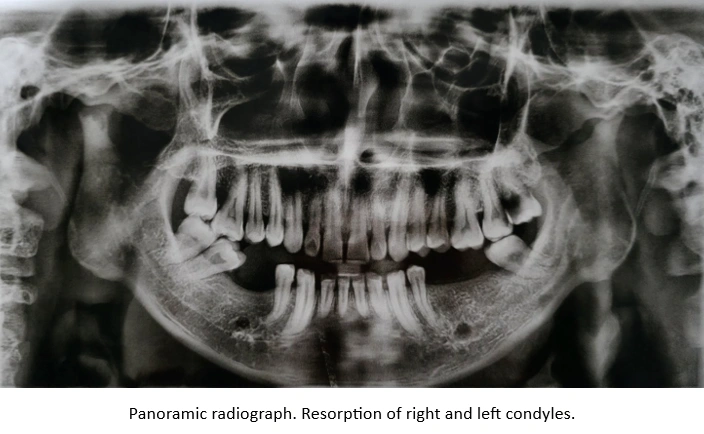
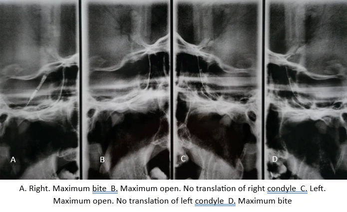
This case presents the non-surgical orthodontic management of a 37-year-old man diagnosed with systemic rheumatoid arthritis, a chronic autoimmune disease affecting multiple synovial joints throughout the body. The patient sought treatment because of persistent pain in both temporomandibular joints (TMJs), limited mouth opening, restricted mandibular movements, and difficulty chewing. Clinical and radiographic examination also revealed TMJ condylar resorption, contributing to compromised mandibular function.
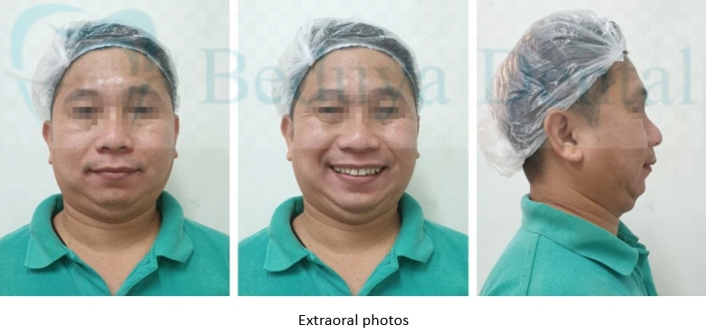
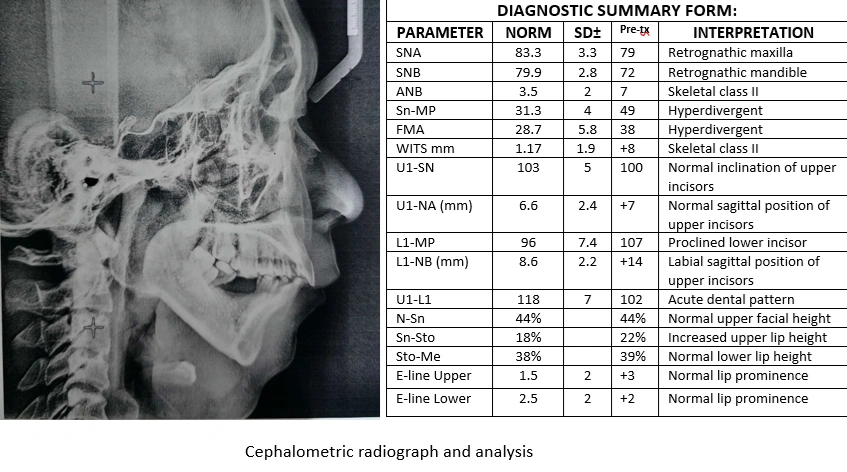
Clinical examination demonstrated a compromised posterior occlusion with multiple missing mandibular molars and mesial tipping of the remaining posterior teeth. Cephalometric analysis demonstrated a Skeletal Class II malocclusion characterized by a retrognathic mandible, posterior mandibular rotation, and a deep bite tendency. These combined skeletal, dental, and joint findings also contributed to a retrusive facial profile with reduced chin prominence.
A non-surgical orthodontic approach was selected to address the skeletal and occlusal problems while avoiding orthognathic surgery. Because the loss of multiple mandibular molars resulted in compromised anchorage, TAD-assisted molar uprighting was performed to reposition the remaining posterior teeth. Periodontally Accelerated Osteogenic Orthodontics (PAOO) was also incorporated to facilitate molar uprighting and accelerate orthodontic tooth movement. Following orthodontic correction, the missing posterior teeth were planned for prosthetic replacement as part of the interdisciplinary treatment sequence.
The patient sought treatment because of persistent pain in both temporomandibular joints, inability to open his mouth normally, and difficulty chewing. He was also concerned about the progressive deterioration of his bite and facial appearance.
The patient had a known history of rheumatoid arthritis and was under the regular care of his physician for ongoing medical management. He reported involvement of multiple peripheral joints, particularly the fingers, wrists, knees, and ankles, with intermittent joint pain, stiffness, and limitation of movement during disease exacerbations. Medical therapy consisted primarily of disease-modifying antirheumatic drugs (DMARDs), with nonsteroidal anti-inflammatory drugs (NSAIDs) and corticosteroids prescribed as clinically indicated for symptom control. He attended regular follow-up appointments with his physician for continued monitoring and adjustment of his medical treatment.
Before seeking orthodontic treatment, there had been no documented evaluation of the temporomandibular joints in relation to his rheumatoid arthritis. The patient also reported no previous history of swelling over either TMJ region.
The patient reported a history of multiple mandibular molar extractions before seeking orthodontic treatment. He stated that he had not worn any removable or fixed prosthesis to replace the missing teeth since their extraction. He also denied any history of significant facial trauma and could not recall any previous injury involving the jaws or temporomandibular joints.
Clinical examination revealed a compromised posterior occlusion with multiple missing mandibular molars and mesially tipped posterior teeth. Cephalometric analysis demonstrated a Skeletal Class II malocclusion characterized by a retrognathic mandible, posterior mandibular rotation, and a deep bite tendency. Both temporomandibular joints exhibited condylar resorption associated with rheumatoid arthritis. These combined skeletal, dental, and joint conditions contributed to TMJ dysfunction, restricted mandibular movements, impaired mastication, and a retrusive facial profile with reduced chin prominence.
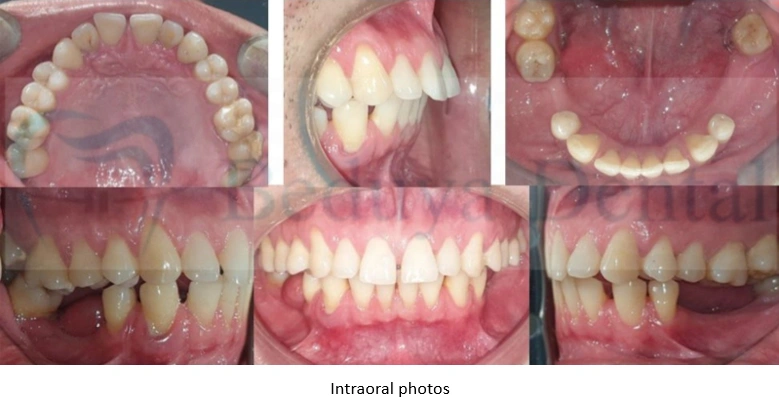
The loss of posterior occlusal support, supraeruption of maxillary first molars and progressive tipping of the remaining mandibular molars further compromised the maxillomandibular relationship, making both orthodontic correction and prosthetic rehabilitation more challenging. Comprehensive diagnosis and interdisciplinary treatment planning were therefore essential before initiating orthodontic therapy.
 Treatment Objectives
The treatment aimed to:
Molar uprighting is the mechanic of choice in this case to re-establish posterior occlusal support following the loss of multiple adjacent mandibular molars. With appropriate biomechanical control, uprighting of the remaining posterior teeth is intended to facilitate anterior mandibular rotation toward a more favorable condylar position while improving the maxillomandibular relationship. This planned mandibular rotation is also intended to increase chin prominence and address the retrusive facial profile.
Because posterior anchorage was compromised by the absence of multiple mandibular molars, temporary anchorage devices (TADs) were strategically placed to provide stable skeletal anchorage for controlled molar uprighting. Partial fixed orthodontic appliances were installed to initiate the planned tooth movement.
Periodontally Accelerated Osteogenic Orthodontics (PAOO) was incorporated into the treatment to facilitate and accelerate molar uprighting. In combination with TAD-supported anchorage, controlled orthodontic forces were applied progressively while periodontal health and tooth mobility were carefully monitored throughout treatment.
Orthodontic treatment was subsequently extended to the maxillary dentition to coordinate the upper and lower arches and establish a more favorable occlusal relationship. A palatal TAD-assisted intrusion system was used to intrude the over-erupted maxillary molars while correcting the buccal inclination of the posterior teeth. These orthodontic corrections were planned to establish appropriate tooth positions and occlusal relationships before proceeding with the prosthetic replacement of the missing teeth.
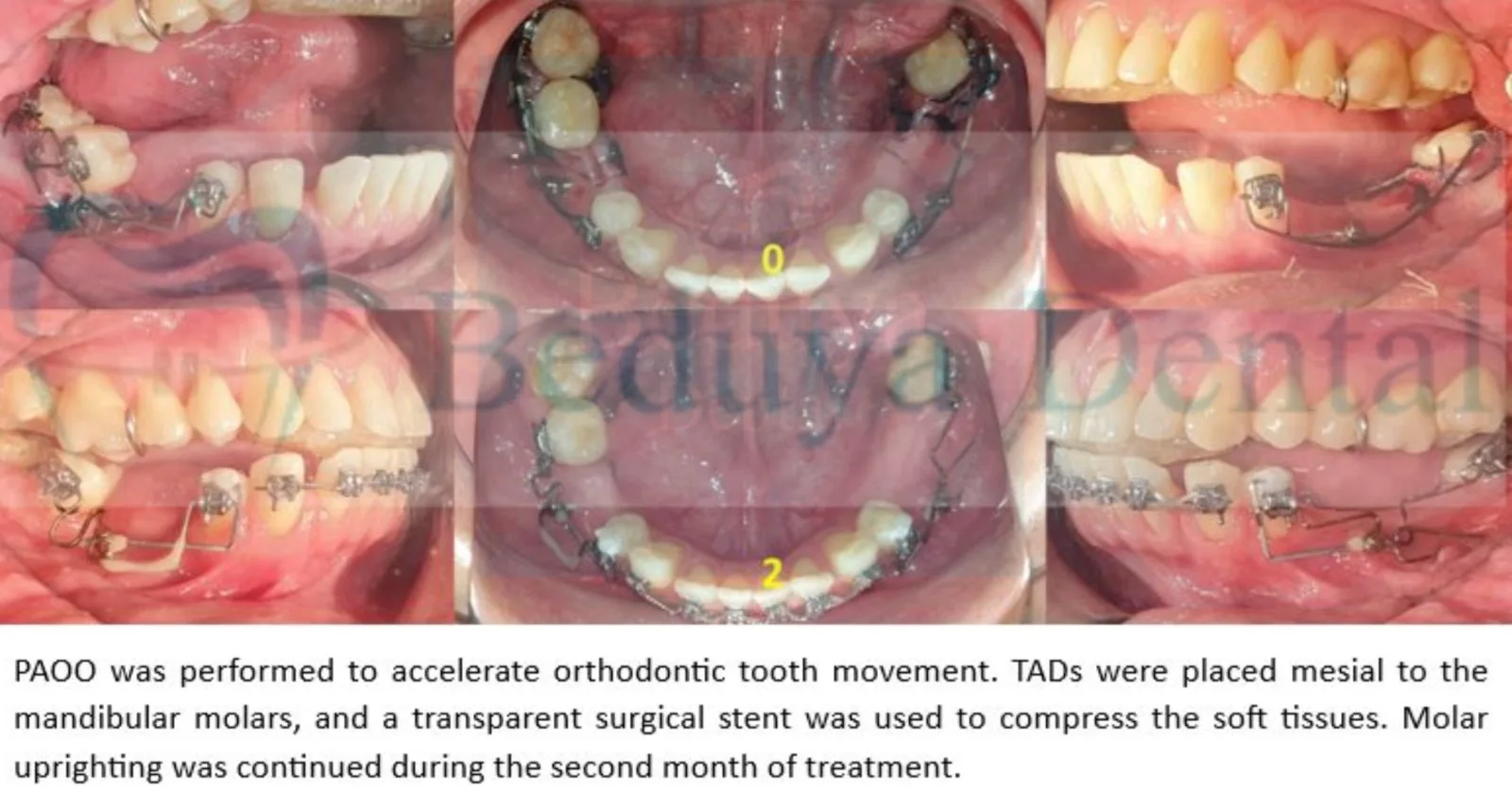
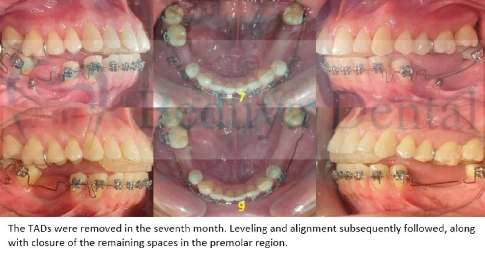
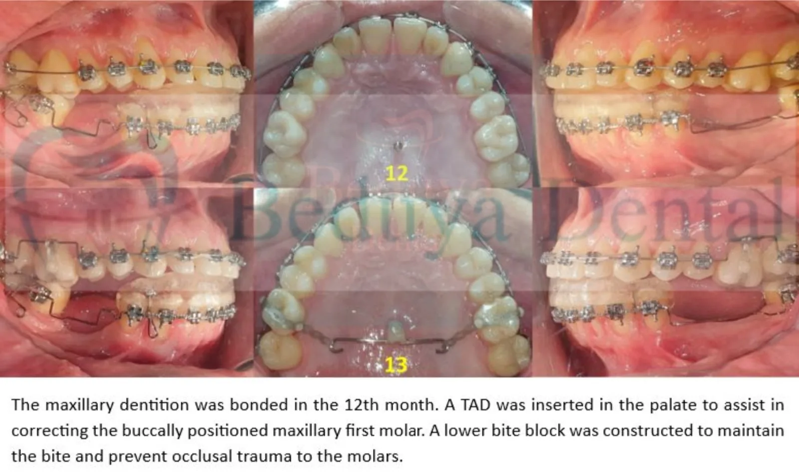
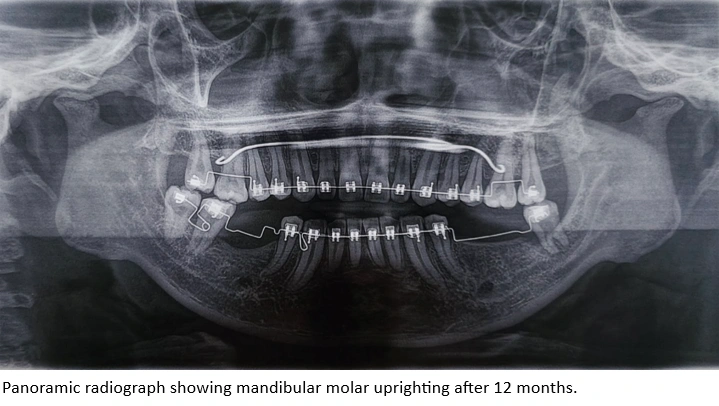
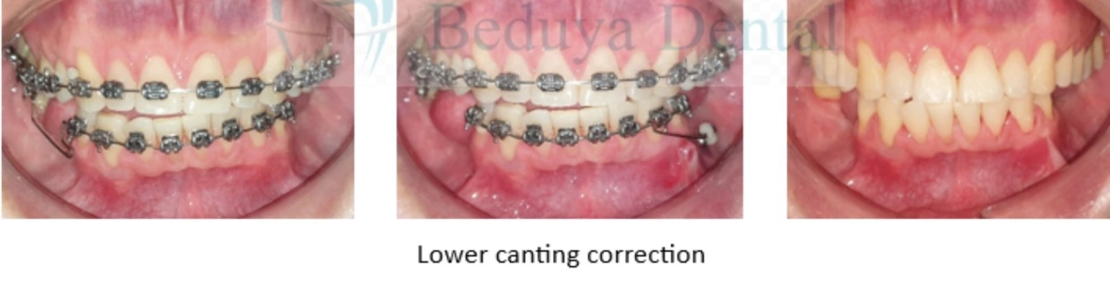
Two interradicular TADs were initially placed in the mandibular arch to assist molar uprighting. Shortly after appliance placement, Periodontally Accelerated Osteogenic Orthodontics (PAOO) was performed to induce the Regional Acceleratory Phenomenon (RAP), facilitating accelerated orthodontic tooth movement during the period of transient bone remodeling. Four months after treatment initiation, panoramic radiographs demonstrated significant simultaneous uprighting of the remaining mandibular molars without evidence of tooth mobility. The mandibular TADs were removed after seven months once adequate molar uprighting had been achieved. Orthodontic treatment then progressed to the maxillary arch, where a palatal TAD-assisted intrusion system was used to intrude the supra-erupted posterior teeth while improving transverse tooth position.
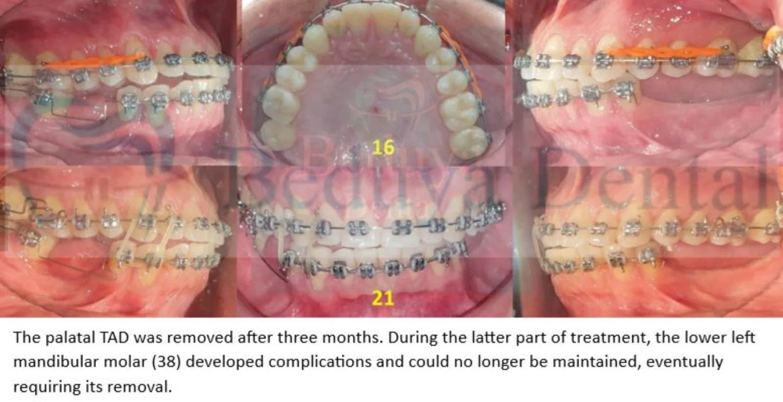
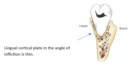 During the later stages of treatment, favorable root torque of the mandibular left third molar could not be fully established during the uprighting process. Severe occlusal wear altered the normal occlusal anatomy of the tooth, making it an unreliable reference for determining its long-axis orientation. As the tooth was uprighted, the root approached the lingual cortical plate in the region of the angle of inflection, compromising its periodontal support. The tooth subsequently developed increased mobility and was eventually extracted.
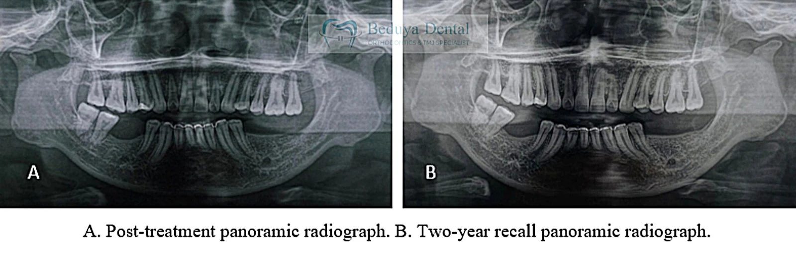
After 12 months of treatment, the remaining mandibular molars were successfully uprighted, establishing a more favorable position for continued orthodontic treatment and eventual restoration of the missing posterior teeth. The panoramic radiograph below demonstrates the improvement in molar position achieved following the uprighting phase.
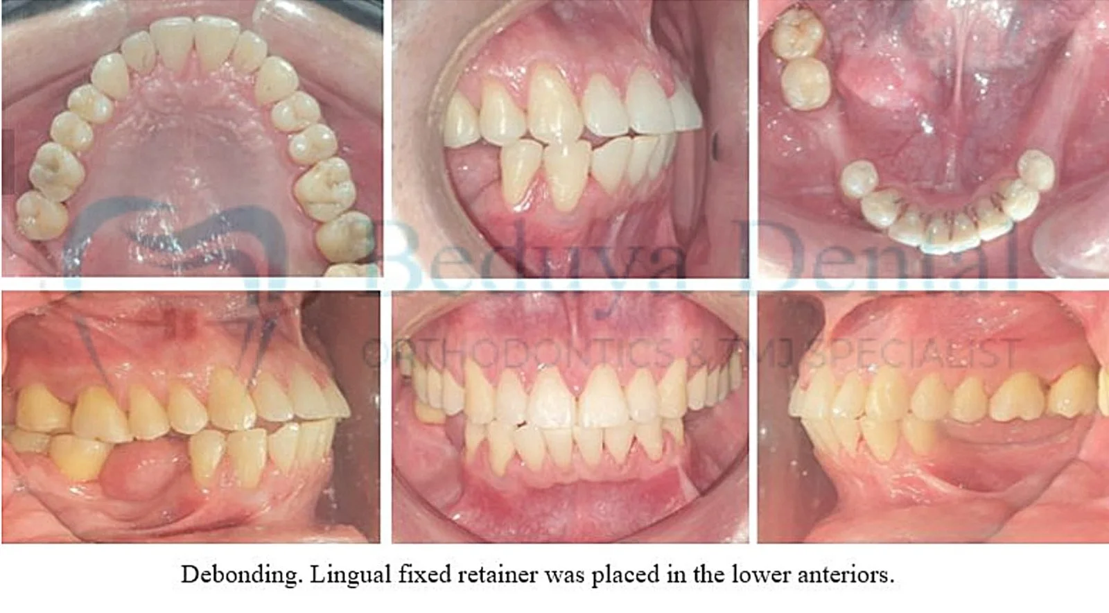
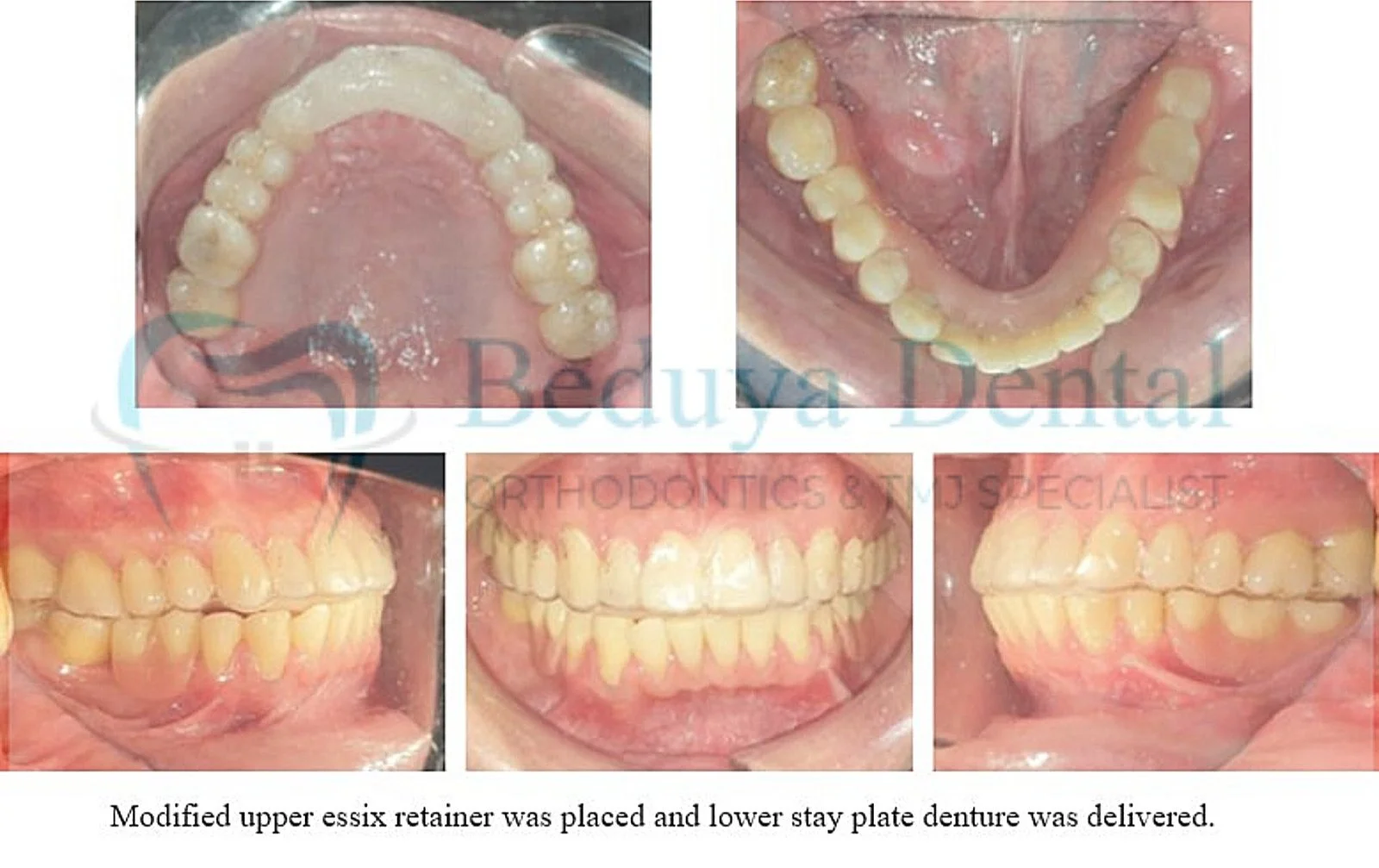
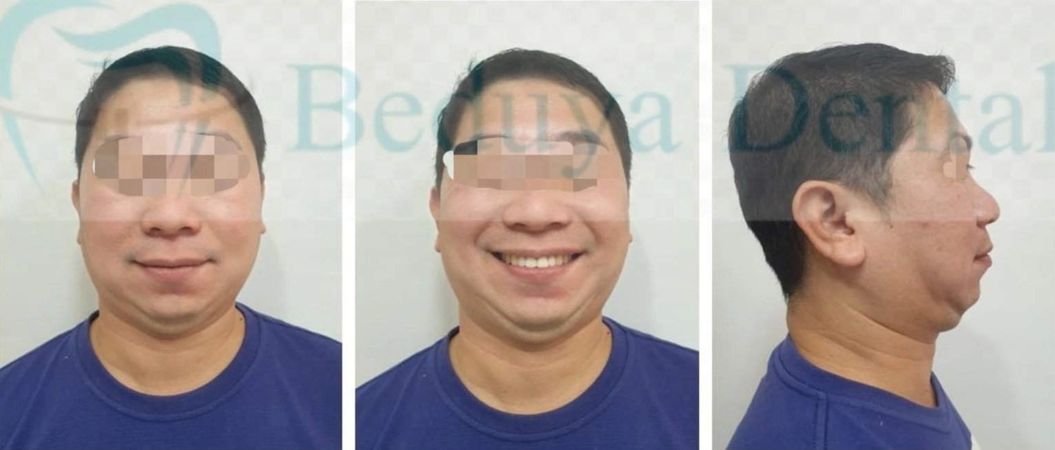
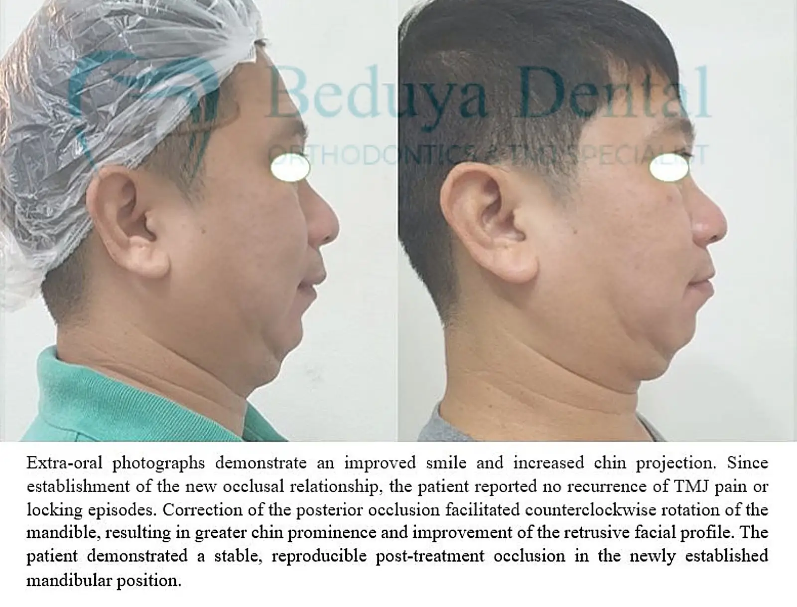
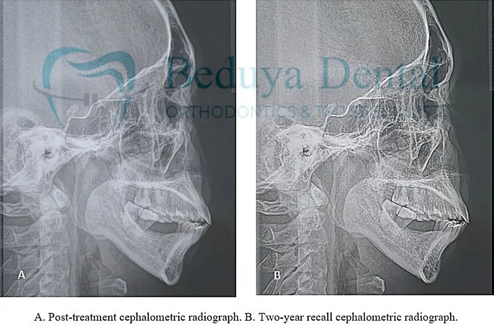
Following completion of orthodontic treatment, the posterior occlusion and overall maxillomandibular relationship were improved. Correction of the posterior occlusion promoted anterior mandibular rotation, resulting in improved chin prominence and a more balanced facial profile.
Functionally, the patient reported complete resolution of TMJ pain. Mouth opening and mandibular movements returned to normal, allowing comfortable mastication and improved daily function. Prosthetic replacement of the missing posterior teeth was subsequently completed to restore posterior support.
Post-treatment photographs demonstrated improved lip posture, a more relaxed mentalis muscle, and enhanced facial balance. The patient’s profile became more harmonious while maintaining a conservative, non-surgical approach.
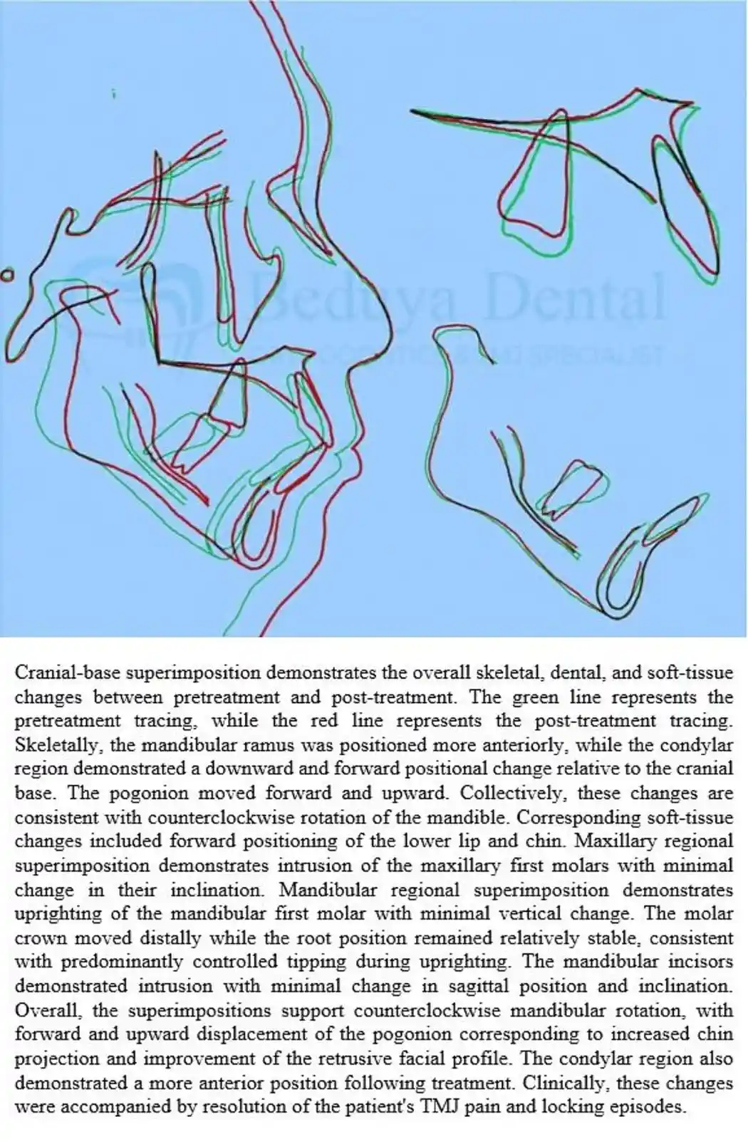 Conclusion
This case demonstrates that Skeletal Class II with a retrognathic mandible and TMJ dysfunction associated with rheumatoid arthritis and TMJ condylar resorption can be successfully managed through a non-surgical orthodontic approach. Strategic use of skeletal anchorage together with PAOO allowed efficient molar uprighting despite compromised posterior anchorage, creating a stable occlusal foundation for definitive prosthetic rehabilitation.
Temporomandibular joint dysfunction is not always caused by the joints alone. In this patient, systemic rheumatoid arthritis contributed to progressive TMJ condylar resorption, while the existing Skeletal Class II malocclusion with a retrognathic mandible, loss of posterior occlusal support, and tipping of the remaining mandibular molars further altered the maxillomandibular relationship and mandibular function. The combined effects of systemic disease, skeletal discrepancy, and occlusal instability likely contributed to the patient’s functional limitations and TMJ symptoms.
Carefully planned orthodontic treatment can play an important role in selected patients by re-establishing posterior occlusal support, improving the maxillomandibular relationship, and promoting a more favorable mandibular position before definitive prosthetic rehabilitation. In dentitions with compromised posterior anchorage, TAD-assisted biomechanics and adjunctive procedures such as Periodontally Accelerated Osteogenic Orthodontics (PAOO) can facilitate controlled tooth movement while minimizing undesirable reciprocal effects.
This case also highlights the importance of three-dimensional root control during molar uprighting. In severely worn posterior teeth, the occlusal anatomy may not accurately represent the long axis of the tooth, making torque control more challenging. CBCT should be considered during treatment planning to evaluate root position within the alveolar housing, particularly in the region of the mandibular angle of inflection, where the lingual cortical plate becomes naturally thinner.
The long-term status of the TMJs remains an important consideration in this case. Although posterior occlusal support was re-established and the patient became asymptomatic following treatment, it cannot be determined from this case whether these changes will provide long-term protection against further TMJ involvement associated with rheumatoid arthritis. Likewise, recurrence of symptoms or progression of condylar resorption cannot be assumed. Continued medical management of rheumatoid arthritis and long-term clinical and radiographic monitoring of the TMJs are therefore important to evaluate the stability of the treatment outcome and any future changes in the joints.
This case reflects the principles of Facial-Driven Multidisciplinary Orthodontics, where orthodontic treatment is planned not only to align teeth but also to restore function, improve facial balance, and coordinate interdisciplinary care for long-term treatment stability.
This clinical case was presented during the scientific lecture “TWIST IT: Exploring the Limits of Orthodontic Tooth Movement with Bone Screws”, delivered at the Association of Philippine Orthodontists. The case was included to demonstrate the application of TAD-assisted molar uprighting and PAOO in managing a complex adult patient with compromised anchorage and TMJ dysfunction.
Disclaimer:
Educational, evidence-informed case reports presented in a patient- and dentist-friendly format. These cases are shared for learning purposes and do not replace individualized professional consultation.
Patient Image & Data Consent Notice
All clinical photographs, radiographs, and records presented in this case report are published with the patient’s informed consent. Identifying information has been removed or minimized to protect patient privacy. These materials are shared solely for educational and professional purposes.