This case highlights the successful correction of occlusal canting in a 51-year-old woman with dentoalveolar collapse caused by the loss of multiple permanent teeth. Dentoalveolar collapse (DAC) is a condition that develops after teeth are lost, causing the remaining teeth to gradually shift into the empty spaces while the surrounding alveolar bone progressively changes. These changes can affect the bite, facial appearance, and the long-term rehabilitation of the dentition (Graber et al., 2017).
To address these problems, a mini-implant-assisted intrusion system was used to reposition the over-erupted upper teeth, correct the occlusal cant, and establish a stable foundation for the patient’s final restorative rehabilitation. This approach restored a more balanced smile, improved oral function, and enhanced facial symmetry.
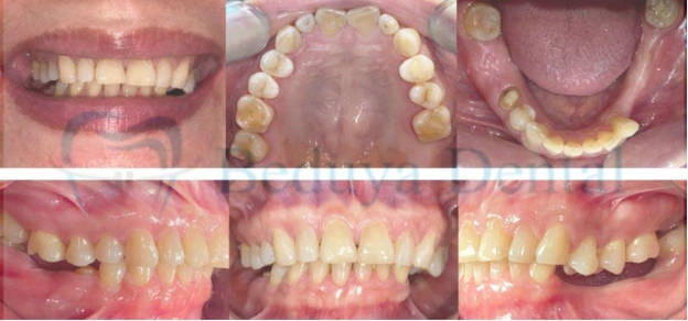
The patient sought treatment because she had difficulty eating after losing several permanent teeth. She was also concerned about the uneven appearance of her smile, which had gradually become more noticeable over time.
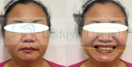
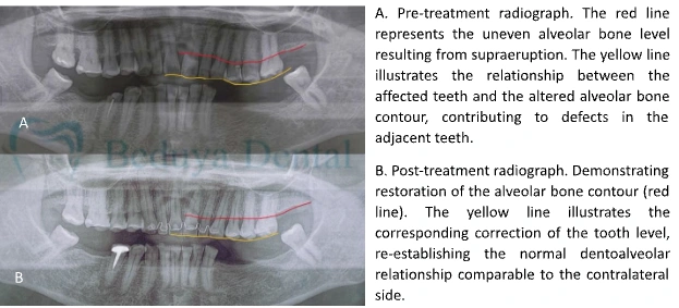
Clinical and radiographic examination revealed dentoalveolar collapse resulting from the loss of multiple permanent teeth. Several upper back teeth had over-erupted into the spaces left by the missing teeth, causing the bite to collapse and the upper teeth to slope downward on the left side. These changes resulted in an uneven smile, facial asymmetry, excessive tooth wear, and reduced chewing efficiency. Simply replacing the missing teeth would not have corrected the underlying problem.
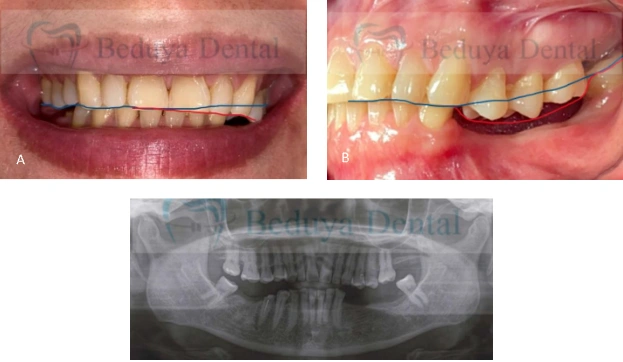
The treatment aimed to:
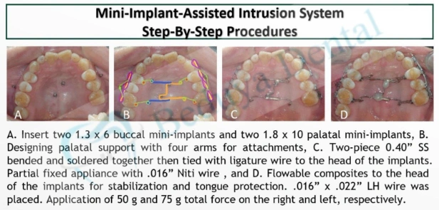
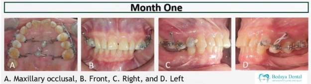
After discussing the available treatment options, the patient chose orthodontic treatment assisted by temporary anchorage devices (TADs). Small orthodontic mini-implants were placed in carefully selected areas of the upper jaw to provide stable support for controlled tooth movement. A custom-designed intrusion system was then used to gradually reposition the over-erupted teeth and level the bite. Partial orthodontic appliances were placed to align the remaining upper teeth before proceeding with the restorative phase of treatment.
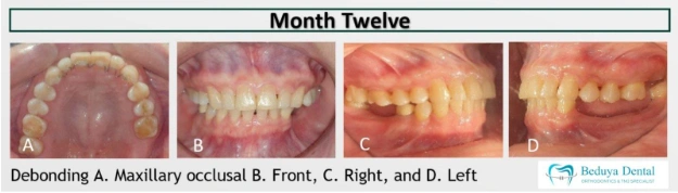
Significant improvement was achieved within the first five months, with successful intrusion of the over-erupted upper teeth and correction of the occlusal cant. The mini-implants were removed after nine months, while the orthodontic appliances were removed after twelve months. Once a healthy and stable bite had been established, restorative treatment was completed to replace the missing teeth and restore proper oral function.
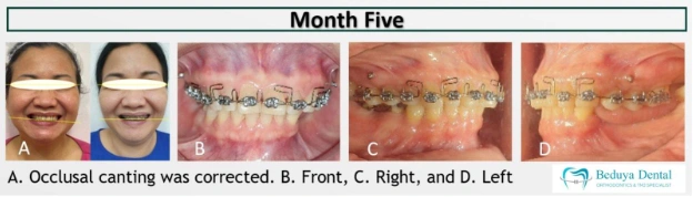
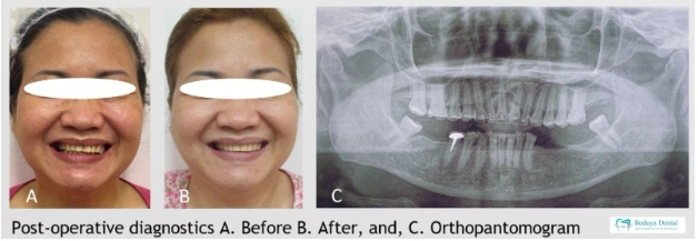
Treatment successfully corrected the uneven bite, improved facial symmetry, lips became symmetrical and restored a balanced smile. Post-treatment radiographs demonstrated good root alignment without evidence of root resorption. By correcting the bite before replacing the missing teeth, the final restorations were completed under more favorable conditions, resulting in improved oral function, facial balance, smile aesthetics, and long-term treatment stability.
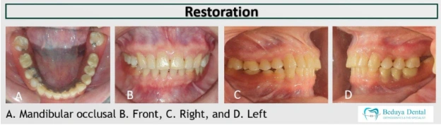
The mini-implant-assisted intrusion system provided a simple, minimally invasive, and predictable solution for correcting occlusal canting associated with dentoalveolar collapse in this adult patient. By re-establishing a functional occlusal plane before restorative care, it created favorable conditions for prosthetic rehabilitation and enhanced both facial harmony and masticatory function. This case demonstrates the value of a coordinated interdisciplinary approach in managing complex adult patients and achieving durable clinical outcomes.
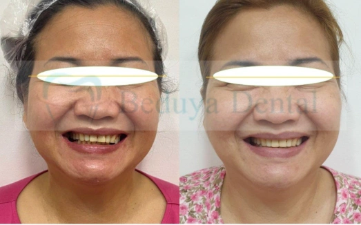
Replacing missing teeth is not always the first step in treatment. In many adults, neighboring teeth gradually drift or over-erupt into the empty spaces, causing the bite to collapse and making restorative treatment more challenging.
In this case, orthodontic treatment was performed first to reposition the remaining teeth and restore a healthy bite before replacing the missing teeth. This carefully planned treatment sequence created a more stable foundation for the final restorations while improving both facial symmetry and oral function.
This case also reflects Dr. Beduya’s philosophy of Facial-Driven Orthodontics, where orthodontic treatment is planned not only to align teeth and correct the bite but also to support facial aesthetics, oral function, and comprehensive dental rehabilitation. Every stage of treatment is carefully coordinated to achieve healthy, functional, and long-lasting results.
This clinical case was presented at the 37th Annual Meeting of the Taiwan Association of Orthodontists (TAO) and the 14th Asia Pacific Orthodontic Congress (APOC), held at TaiNEX 2, Taipei, Taiwan, on December 3–5, 2024.
Azami N, Nanda R, Uribe F. Effective Vertical Control of the Entire Maxillary Arch with a Palatal TAD-Supported Appliance. Journal of Clinical Orthodontics. 2020;54(10):620-629.
Chang CJ, Lin WC, Chen MY, Chang HC. Evaluation of Total Bone and Cortical Bone Thickness of the Palate for Temporary Anchorage Device Insertion. Journal of Dental Sciences. 2021;16:636-642.
Beduya, E.K.S. Correction of Occlusal Canting in an Adult with Dentoalveolar Collapse Using a Mini-Implant-Assisted Intrusion System: A Case Report. 37th Annual Meeting of the Taiwan Association of Orthodontists (TAO) and the 14th Asia Pacific Orthodontic Congress (APOC), Taipei, Taiwan, December 3–5, 2024.
Graber LW, Vanarsdall RL Jr, Vig KWL, Huang GJ, eds. Orthodontics: Current Principles and Techniques. 6th ed. St. Louis, MO: Elsevier; 2017.
Disclaimer:
Educational, evidence-informed case reports presented in a patient- and dentist-friendly format. These cases are shared for learning purposes and do not replace individualized professional consultation.
Patient Image & Data Consent Notice
All clinical photographs, radiographs, and records presented in this case report are published with the patient’s informed consent. Identifying information has been removed or minimized to protect patient privacy. These materials are shared solely for educational and professional purposes.